Prof. Dr. Selçuk Palaoğlu
Beyin Ve Sinir Cerrahisi
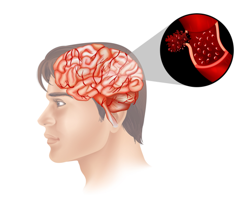
Beyincik sarkmasının tedavisi vardır. Öncelikle ameliyat edilmesi gereken olgular titizlikle seçilmelidirler. Bir çok kişide manyetik rezonans görüntülemesinde beyincik sarkması rapor edilebilir. Ancak gerçekte çok az bir kısmında ameliyata gereksinimi vardır. Tedavi edilmesi gereken olgularda ameliyat tek çözümdür. Ameliyatın hedefi sarkan beyincik kısmının omurilik başlangıcına yaptığı baskıyı azaltmaktır. Ameliyat yeri beyincik, beyin sapı ve omurilik buluşma yerindedir. Mikroskop, mikroşirürji ameliyatın temel altyapılarıdır. Ameliyata omurilik ve beyin sapını rahatlatacak kemik yapıların alınması ile başlanır. Bazen sadece kemik yapıların alınması bile yeterli olabilir. Ameliyatın bundan sonraki sürecinde beyincik ve omurilik zarları açılır. Dış zar olan duranın açılması yeterli olabilir. Ancak beyin omurilik sıvısı dolanımının sağlanamadığı görülürse iç zar olan araknoidde açılabilir. Bazı olgularda işlevsiz olduğu düşünülen sarkmış beyincik kısmı çıkartılabilir. Ameliyatın son kısmında bedenden alınmış veya sentetik bir yama ile duraplasti denilen işlemle açılan dura geniş bir biçimde kapatılır. Ameliyatın amacı beyinciğin sarkması sonucu gelişen beyin sapı ve omurilik basısına bağlı nörolojik kötüleşmeleri ve beyin omurilik sıvısı dolanım bozukluğunu düzeltmektir. Yapılan ameliyatın adı foramen magnum dekompresyonu ve “sisterna magna remodelling” dir.
Chiari malformation surgery
A Chiari malformation, previously called an Arnold-Chiari malformation, is where the lower part of the cerebellum and brain stem pushes down into the spinal canal. Surgical treatment of Chiari malformation is available. Firstly, the cases that need to be operated must be selected carefully. In most people, a Chiari malformation may be reported in magnetic resonance imaging. However, in reality, only a small number of patients need surgery. Surgery is the only solution in cases that need to be treated. The aim of the surgery is to reduce the pressure on the cervical cord due to slumped cerebellum. The area of operation is at the junction of the cerebellum, brain stem and spinal cord. Microscopy and microsurgery are the basic infrastructures of the surgery. Surgery is started with the removal of bone structures that will relax the spinal cord and brain stem. Sometimes it is enough to take only bone structures. The cerebellum and spinal cord membranes are opened after the operation. The opening of the outer membrane can be sufficient. However, if the cerebrospinal fluid circulation is not followed, it can be opened in the inner membrane called as arachnoid. In some cases, the downward displacement of cerebellum part - tonsillary, which is considered to be dysfunctional, can be removed. At the end of the operation, the dura,is closed either with soft tssue taken from the body, or dural patch. The aim of the surgery is to correct the neurological worsening due to the anomaly of the cerebellum and to correct the cerebrospinal fluid discontunity. Surgical procedure is foramen magnum decompression and cisterna magna remodeling.
Bu makalenin DoktorTakvimi web sitesinde yayımlanması, yazarın açık izniyle yapılmaktadır. Web sitesindeki tüm içerikler, fikri ve sınai mülkiyet mevzuatı kapsamında uygun şekilde korunmaktadır.
DocPlanner Teknoloji A.Ş. web sitesi tıbbi tavsiye sunmaz. Bu sayfanın içeriği, metinler, grafikler, görseller ve diğer materyaller de dahil olmak üzere, yalnızca bilgilendirme amacıyla oluşturulmuştur ve tıbbi tavsiye, teşhis veya tedavinin yerini almak amacı taşımaz. Herhangi bir sağlık sorununuzla ilgili şüpheniz varsa, bir uzmana danışınız.